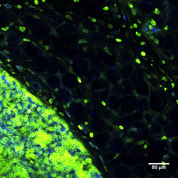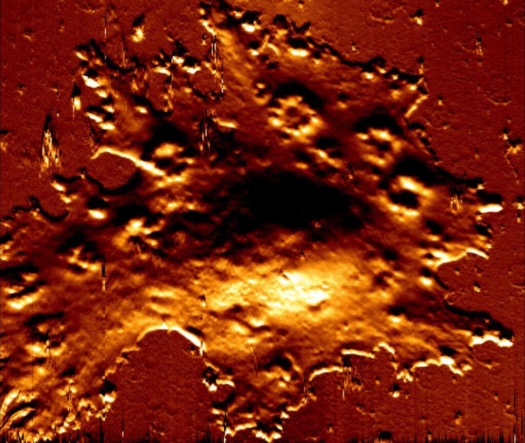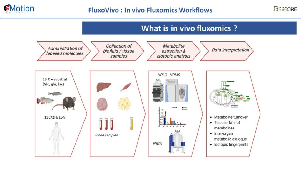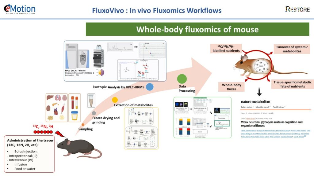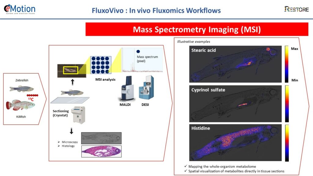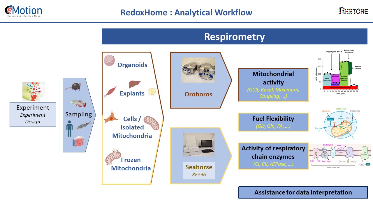
Data Management, Processing and Analysis
In the life cycle of a biological data, which goes from its acquisition to the formatting of results, the processing and analysis constitute a key step to making sens of the data. Analysis uses a set of mathematical and statistical operations whose goal is to improve the quality of the data and to extract relevant information.
Whether the data is acquired on or off site, the data management and analysis platform offers a set of skills and makes available to users computing and software resources covering the fields of processing, analysis, instrumental development and mathematics/statistics. It is therefore possible to propose targeted offers ranging from the reconstruction of raw data to their exploitation. Depending on the user’s request, dedicated staff will evaluate the needs and propose an adapted service ranging from support to autonomy.
EXPERTISE
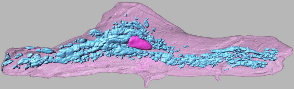
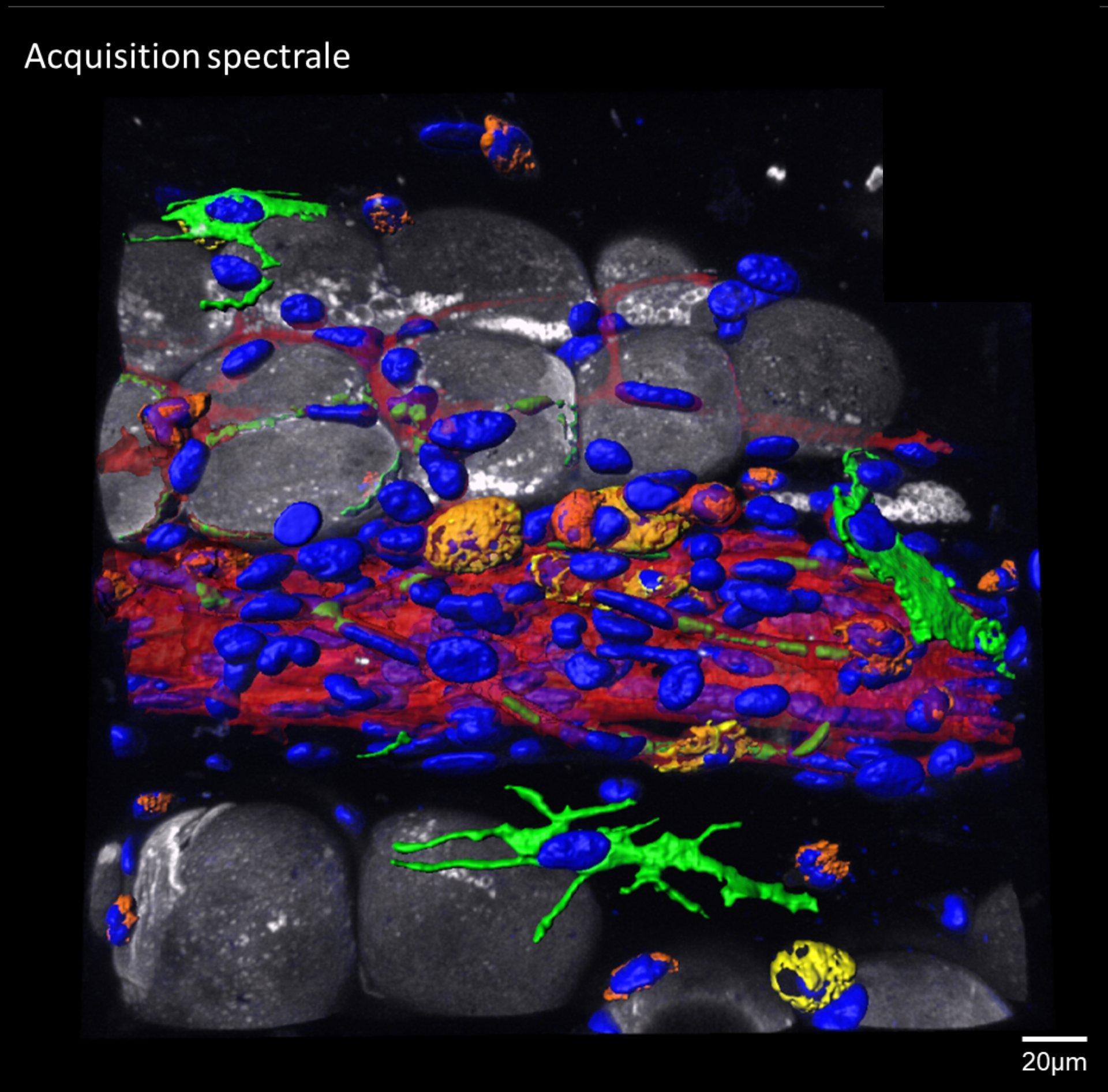
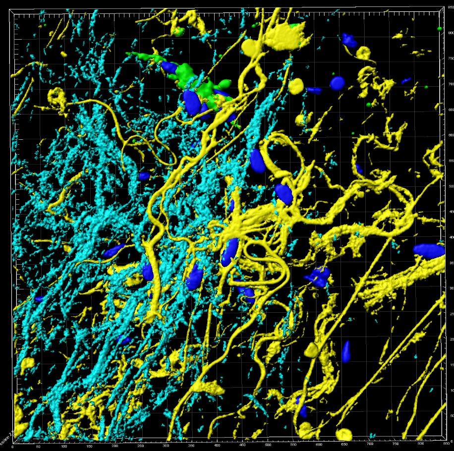
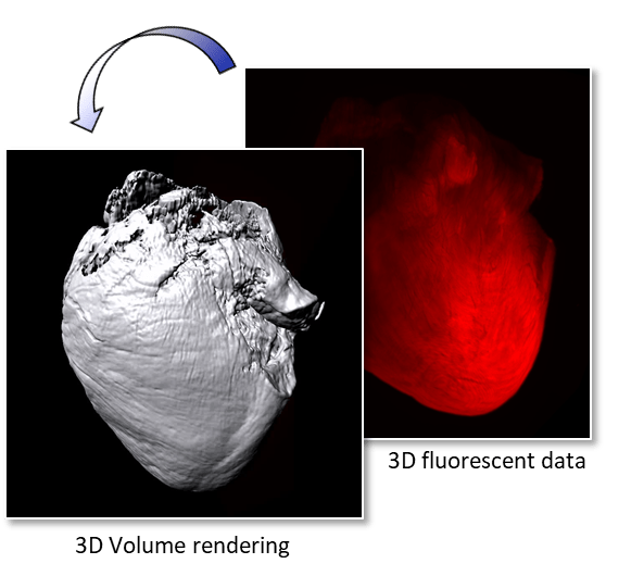
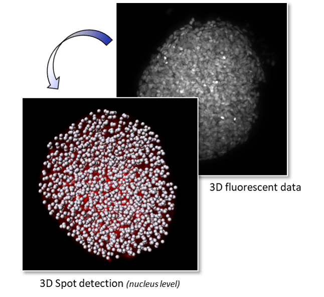
- 3D modeling of living tissues: Imaris & Amira
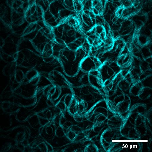
- In depth 3D time resolved image processing: Fiji & Icy
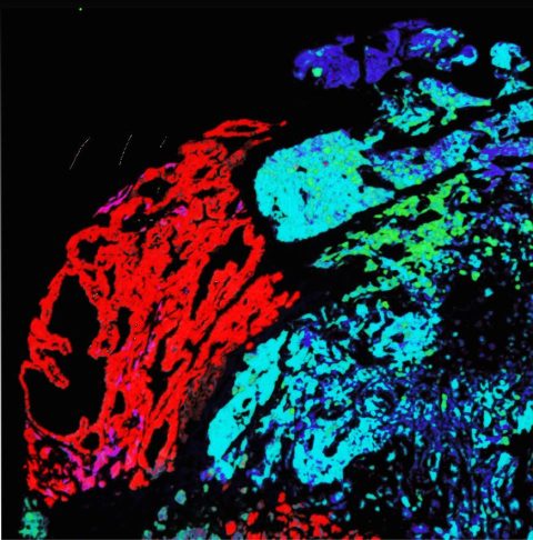
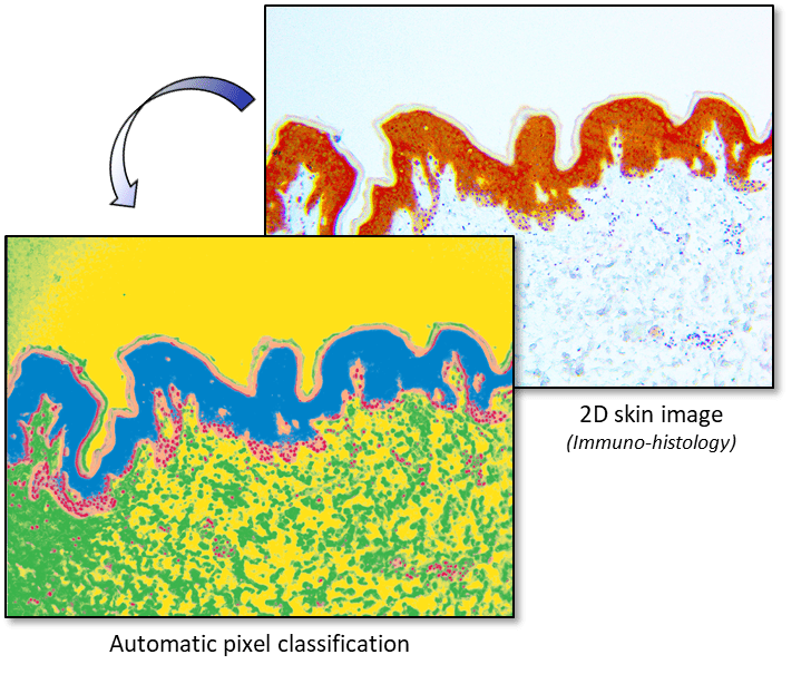
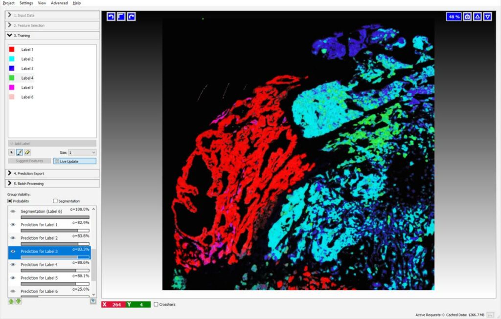
- Automated pixel & object classification: Ilastik, Vision4D & Matlab
- Time-resolved object tracking on 2D or 3D images of living samples: Imaris & Fiji
- Automation for high volume data processing: Matlab & Fiji
- Statistical analysis of biological data: GraphPad Prism
- Development of graphical interfaces for data visualization and manipulation: Matlab
- High throughput and high information content analysis: HCS Studio & Harmony
- Development of databases for heterogeneous data management: Matlab, Sql, Php
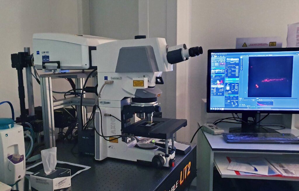
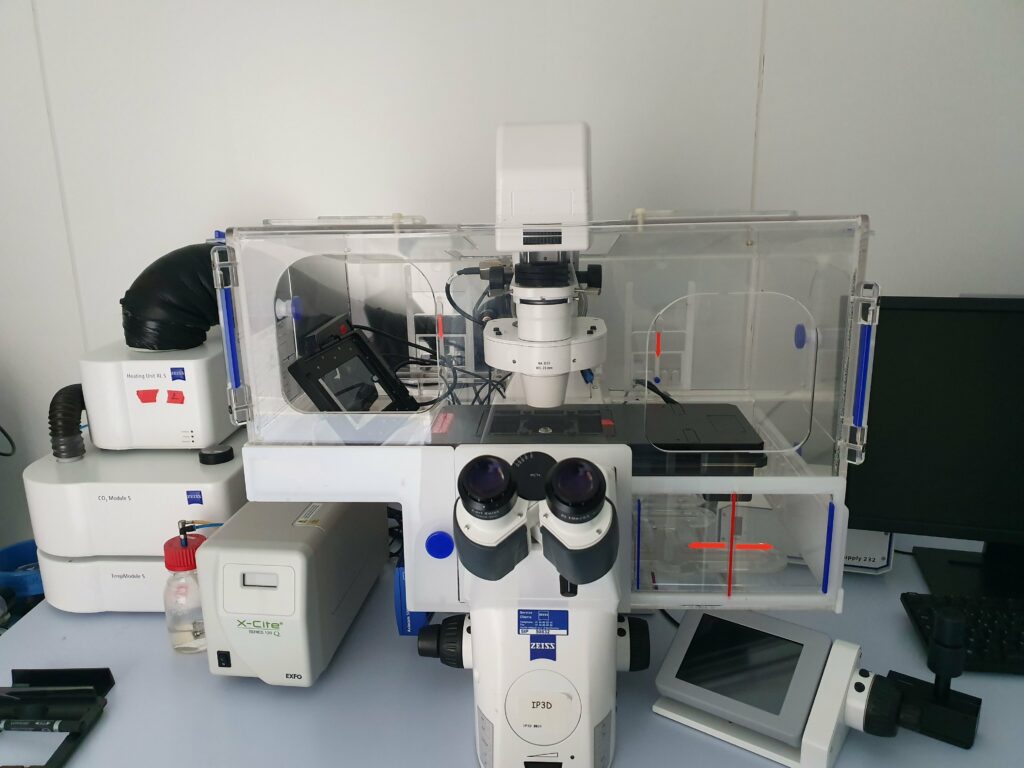
EQUIPEMENTS
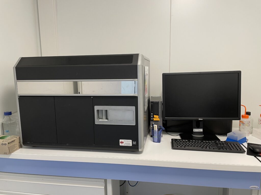
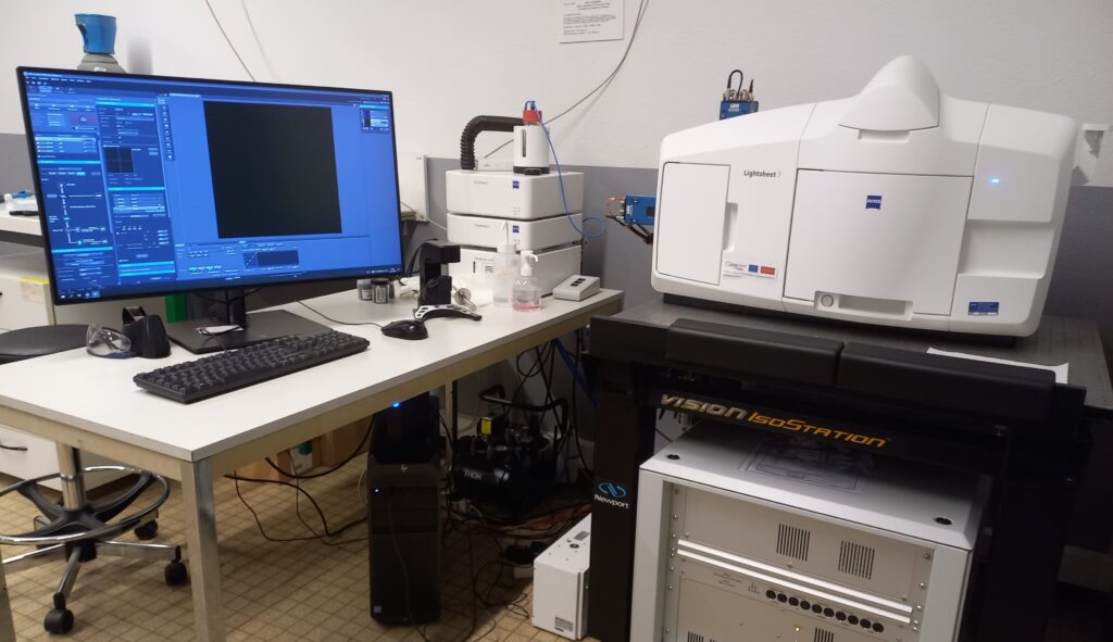
The platform relies on a set of analysis stations with sufficient computing power and high-performance graphics cards for the analysis of large volumes of data, the modeling and the visualization of very high-resolution 3D & 4D images using the most powerful software available at the moment.
3 Imaris (Bitplane) licences
- 3D & 4D modeling, 2D & 3D tracking
2 Amira (ThermoFisher) licences
- 3D modeling, 3D & 4D image processing
1 Vision 4D (Arivis) licence
- 3D & 4D modeling, pixel & object classification
CONTACT

The platform's engineer evaluates the user's request and proposes the most suitable solution to his needs:
- Advice and support of the user at the various stages of data processing and analysis
- Hardware and software use support
- Software training until users become autonomous
- Development on request of specific scripts for the processing and analysis of large volumes of data
- Support in the implementation of research and development projects in the form of collaboration